Basics: Image Orientation, Image Interpretation, Ultrasound Terminology, Machine Settings according to the case and Usage of Different Knobs, Different Types of Probes and their Selection according to the Scan, Artifacts (false pictures that may be misinterpreted as pathologies) and their Remedies, Probe Handling and Focussing, Interventional Orientation.
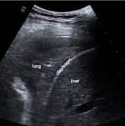
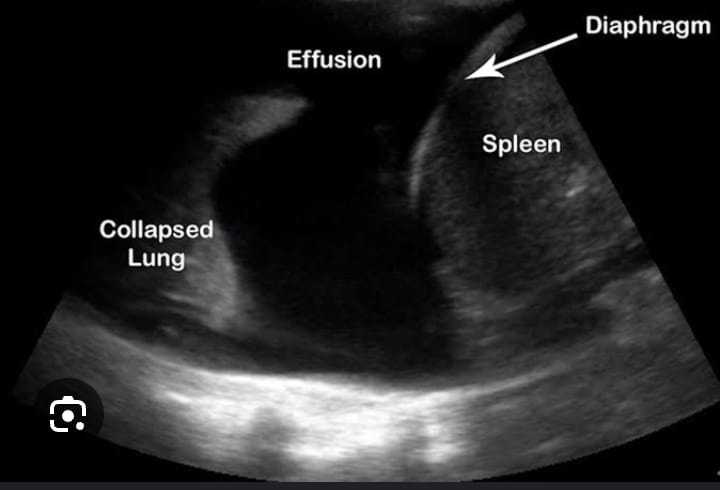
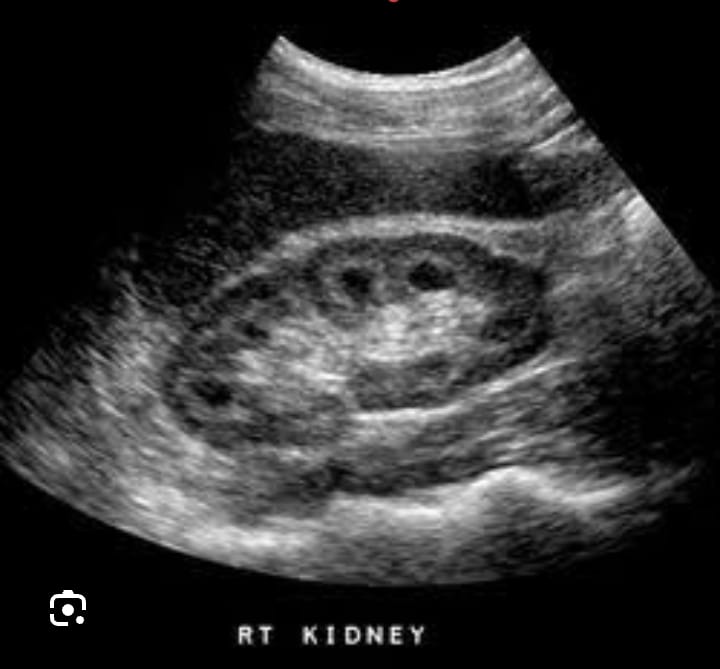
Abdomen: Liver, Gall Bladder, Bile Ducts, Pancreas, Kidney, Bladder, Prostate, Aorta, Abdo Wall, Peritoneum, Chest, Appendix, Retroperitoneum, Breast.
Obstetrics & Gynaecology: Transvaginal scanning is an integral part of the course.
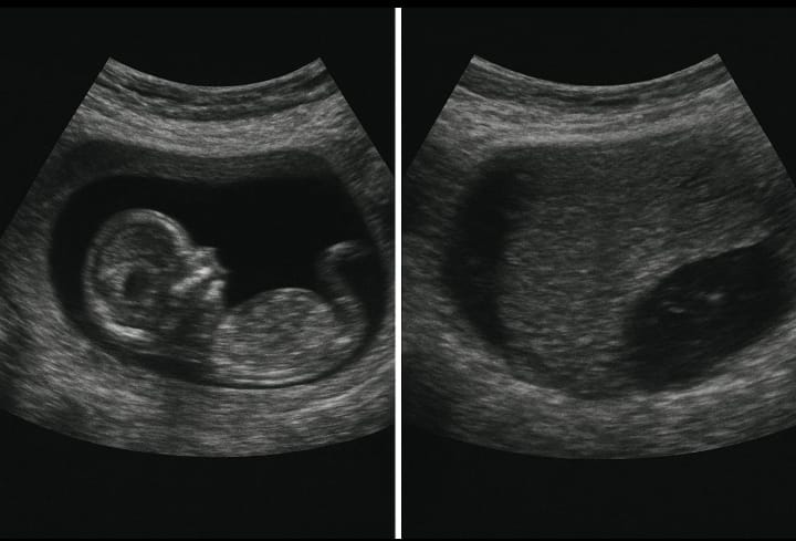
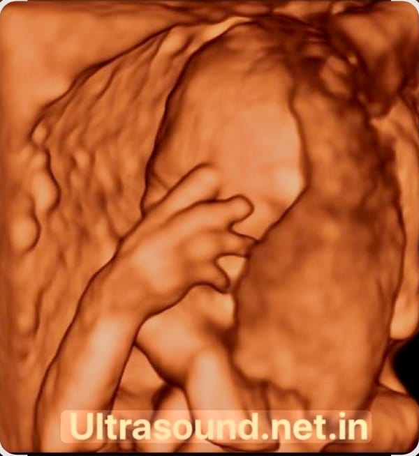
First Trimester, Ectopic, Fetal gestational age estimation, Placenta, Multiple pregnancy, Hydrops, Normal & abnormal fetal anatomy including Gross Congenital Anomalies, Scar Thickness, Cervical Incompetence, IUD, Postpartum ultrasound
Normal female pelvic anatomy and variations Uterine abnormalities- Structural, endometrial, myometrial, Ovarian & Adnexal pathologies, Infertility assessment
Basics:Image Orientation, Image Interpretation, Ultrasound Terminology, Machine Settings according to the case and Usage of Different Knobs, Different Types Of Probes and their Selection according to the Scan, Artifacts (false pictures that may be misinterpreted as pathologies) and their Remedies, Probe Handling and Focussing, Interventional Orientation.
Obstetrics & Gynaecology: Transvaginal scanning is an integral part of the course.
First Trimester, Ectopics, Fetal gestational age estimation, Placenta,Multiple pregnancy, Hydrops, Normal & abnormal fetal anatomy including Gross Congenital Anomalies,ScarThickness, Cervical Incompetence, IUD, Postpartum ultrasound
Basics:Image Orientation, Image Interpretation, Ultrasound Terminology, Machine Settings according to the case and Usage of Different Knobs, Different Types Of Probes and their Selection according to the Scan, Artifacts (false pictures that may be misinterpreted as pathologies) and their Remedies, Probe Handling and Focussing, Interventional Orientation.
Abdomen:Liver,Gall Bladder, Bile Ducts,Pancreas,Kidney,Bladder,Prostate,Aorta, Abdo Wall,Peritoneum, Chest, Appendix, Retroperitoneum, Breast
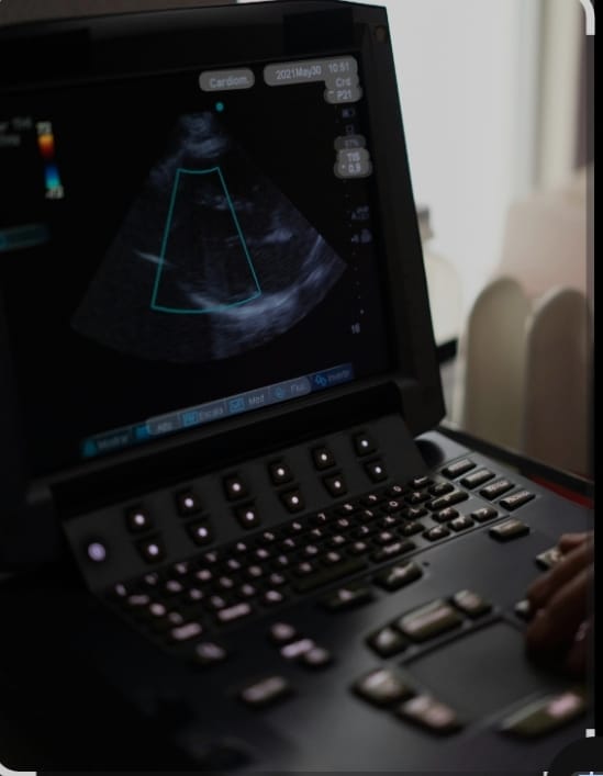
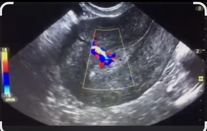
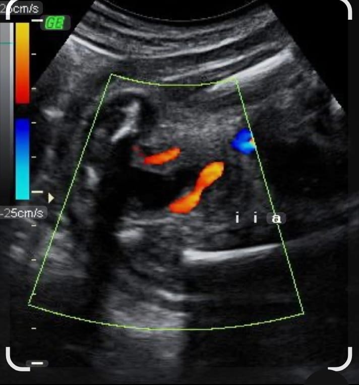
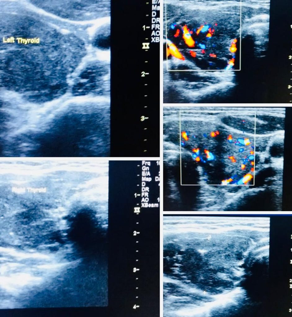
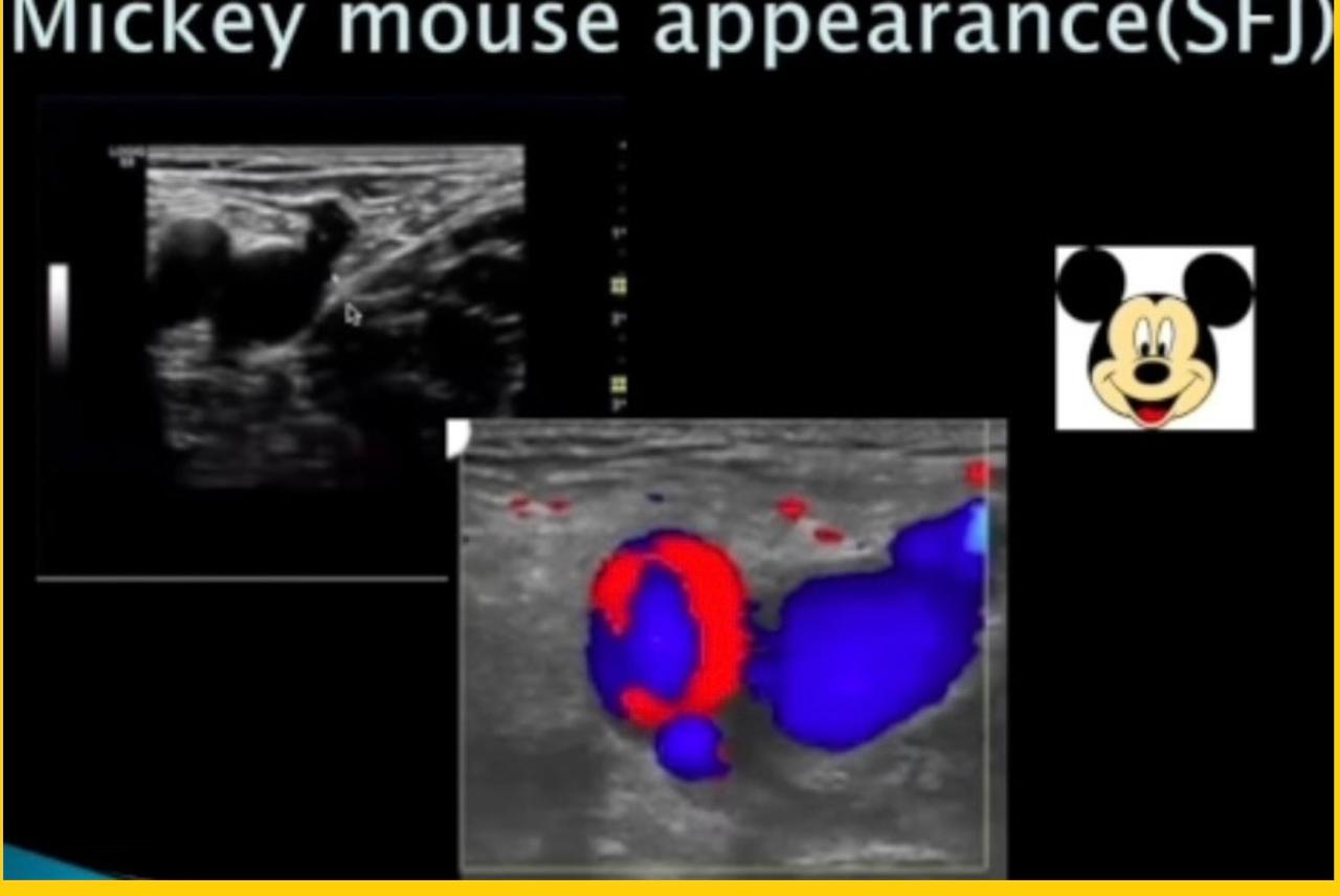
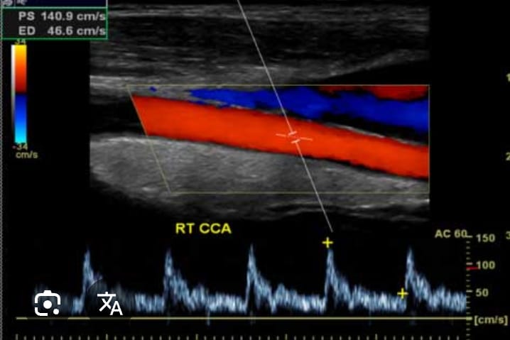
Topics Covered: Basic Physics & Instrumentation, Adjustment of the Machine to get Optimum Picture in Spectral Doppler, Color Flow, Power Doppler, Abdominal Doppler, Peripheral Vascular Doppler, Carotid Doppler, Thyroid, Scrotal, Penile Doppler, Doppler in OBS – GYNAE.
Basics of ultrasound including transvaginal,Normal female pelvic anatomy,Uterine ovarian and adenexal pathologies ,Follicular monitoring,Ovulation disorders,Sonosalpingography
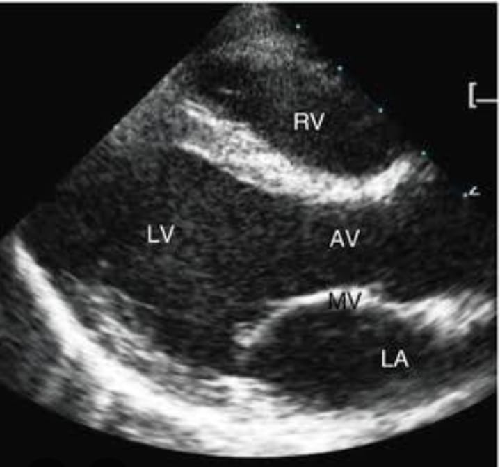
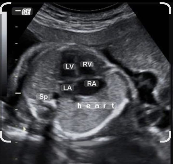
Basic Physics & Instrumentation, Scanning protocols, Adjustment of the machine to get optimum picture,Various views & windows, normal and abnormal fetal cardiac anatomy, detailed study of all congenital cardiac anomalies